What is a Hip Impingement?
Hip, or femoroacetabular impingement (FAI) is a condition where the bones of the hip are abnormally shaped. Because they do not fit together perfectly, the hip bones jam or rub against the soft tissue structures of the joint. This can cause pain and loss of function. FAI is now recognised as a significant cause of premature hip arthritis.
What is hip impingement?
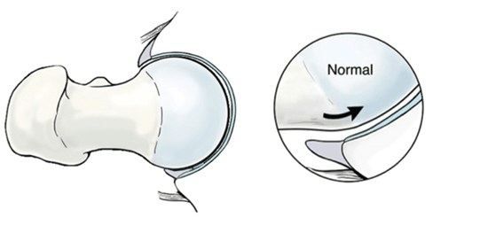
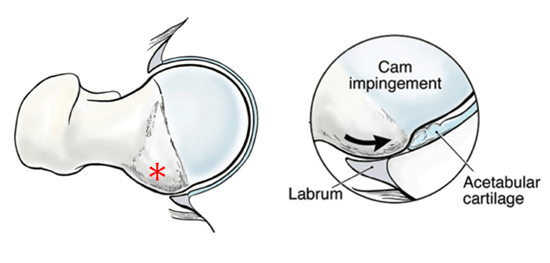
Femoroacetabular impingement (FAI) usually occurs because the hip does not form as a perfect ball and socket during the growing years. This includes “cam” and “pincer” forms of FAI shown below. Often the FAI is a combination of both cam and pincer impingement.
FAI leads to the cartilage and labrum being pinched or squeezed between the abnormal ball and socket when the hip rotates, limiting movement.
Over time or through a traumatic event, this action can result in the tearing of the labrum and breakdown of the articular cartilage. This can cause pain and loss of function. Over time, the progression of this damage can lead to premature arthritis in the hip joint.
What are the symptoms of hip impingement?
People with impingement usually have pain in the groin area, although it can be felt elsewhere. Sharp stabbing pain may occur with turning, twisting, and squatting, or specific sporting activities, but sometimes, it is just a dull ache. Catching, clicking or popping may indicate a labral tear associated with the impingement.
When symptoms first occur, it is helpful to try and identify an activity or something you may have done that could have caused the pain. Sometimes, you can just back off on your activities, let your hip rest, and see if the pain will settle down. Over-the-counter anti-inflammatory medicines (ibuprofen, naproxen) may help.
Many people with the x-ray appearances of FAI never get significant problems, probably because they are able to avoid pushing their hip into positions of impingement. However people with large abnormalities or athletically active people, who work the hip joint more vigorously, may begin to experience pain earlier than those who are less active.
How is impingement diagnosed?
A diagnosis of impingement is suggested by the pattern of hip pain and limitations. Other problems can mimic hip impingement, including hernia, groin muscle injuries and back problems. An examination will help to exclude these diagnoses.
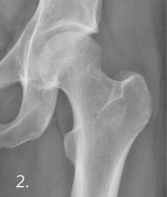
X-rays: of your hip will be performed to assess the shape and orientation of the bones.
MRI scan: will show up labral tears and cartilage damage.
Local anaesthetic: is usually injected into the hip at the same time as the MRI scan. This is important to figure out if your pain is truly coming from within the hip joint itself.
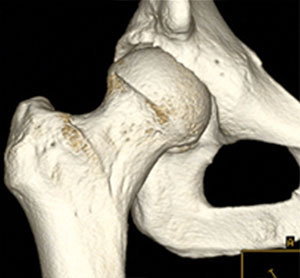
CT scan: In some patients, if we are considering surgery, we will perform a 3D CT scan to accurately map the shape and size of the abnormalities we need to remove during surgery. If you have early arthritis, CT is important to ensure you have enough cartilage in the joint to make hip arthroscopy worthwhile.
CT scan of hip before and after impingement surgery to remove cam bump (white circle) from femur
How is impingement treated?
The initial treatment of impingement is non-surgical. �This is a combination of:
- Activity modification to avoid painful movements and activities
- Anti-inflammatory medications
- Physiotherapy to improve the range of motion in your hip and strengthen the muscles that support the joint. This can relieve some stress on the injured labrum or cartilage.
- Steroid injection: If the hip is particularly irritable and inflamed, a steroid injection may be used to help settle the inflammation.
Surgical treatment
Surgical treatment is indicated if:
- You have tried non-operative treatment and it hasn’t improved your symptoms enough.
- Scans show that the impingement is already causing damage to the joint surfaces or labrum
The most common types of impingement can be treated arthroscopically, that is via small (1cm long) incisions on the side and front of the thigh. In some cases, the deformity is too severe to be managed arthroscopically and you will be referred to see a pelvic surgeon for a different type of operation.